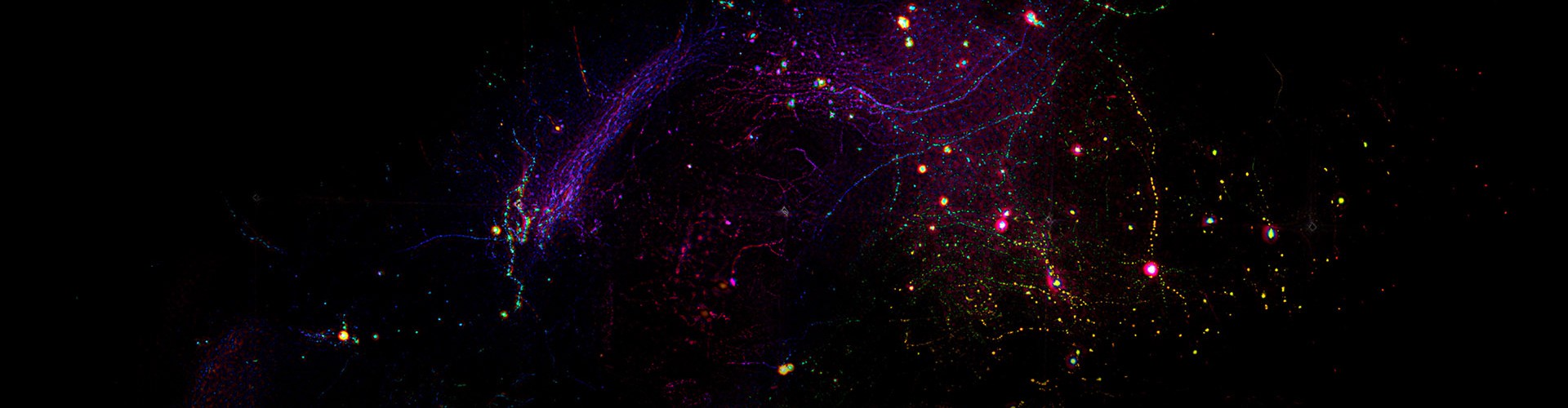
Our laboratory's mission to attain a nuanced appreciation of physiological and pathological phenomena hinges on the ability to perform non-invasive, three-dimensional imaging across opaque tissues. This approach spans various spatial and temporal dimensions, thereby revealing the complex tapestry of intercellular interactions at the organ level. Despite its potential, this methodology faces formidable obstacles such as vast data throughput requirements, optical heterogeneity, surface irregularity, and phototoxic effects. These challenges necessitate a delicate balance between volume size, resolution, speed, sample vitality, and system complexity.
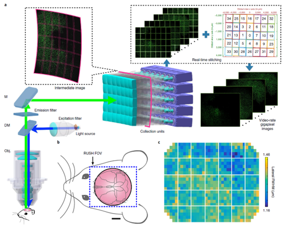
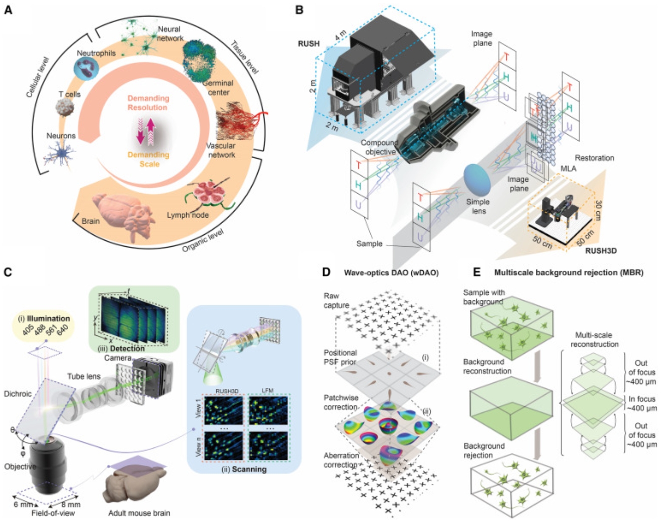
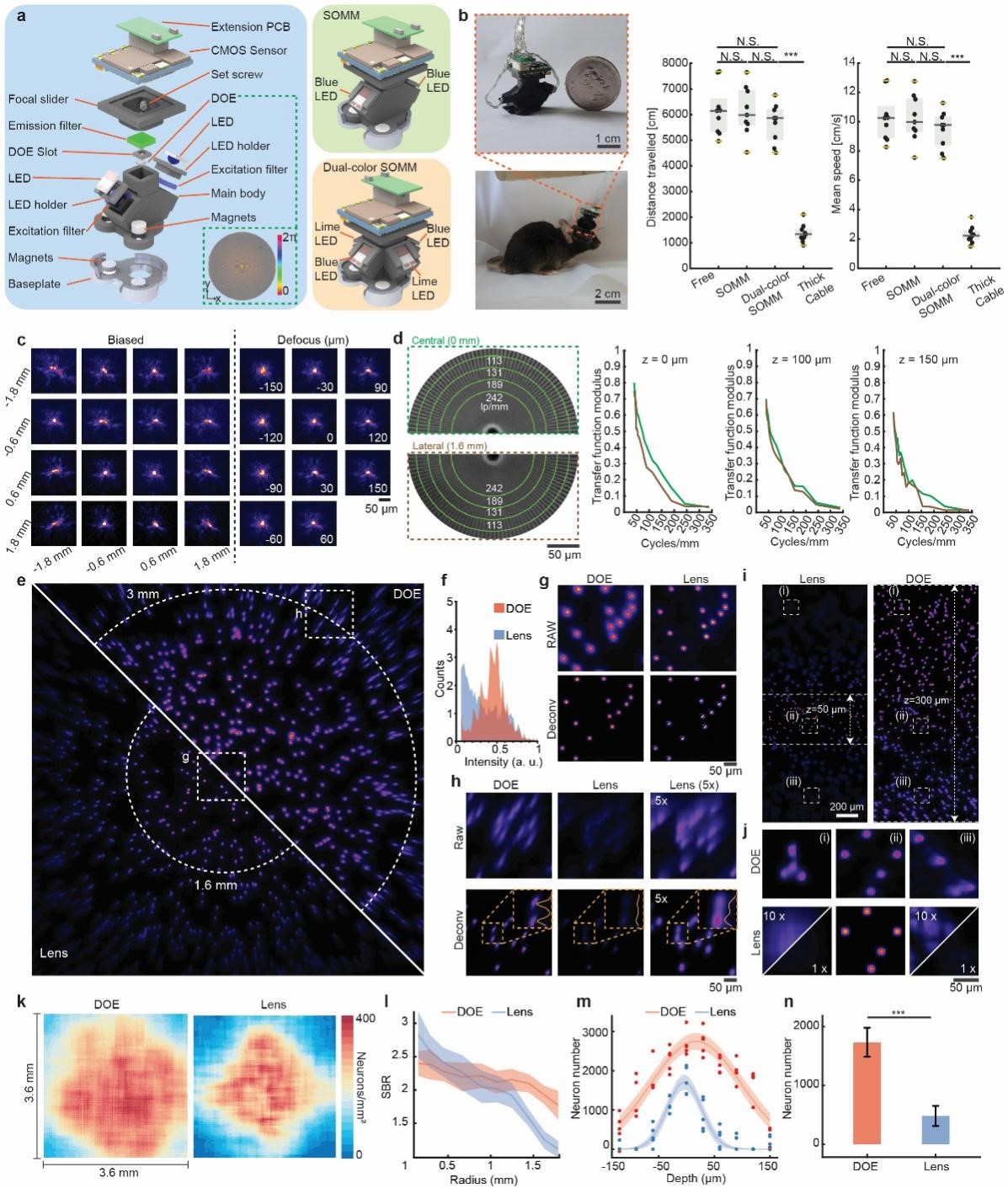
To surmount these barriers, we have integrated advances in computational optics, biological engineering, and machine learning to develop a suite of pioneering tools that enhance the capacity of intravital imaging. Notably, our RUSH technology achieves over a centimeter field of view (FOV) with single-cell resolution. The RUSH3D variant extends these capabilities into three-dimensional space, while RUSH-mini condenses this technology into a compact form factor weighing less than three grams, suitable for studies involving freely moving animals.
These innovations find wide applicability in the fields of neuroscience, immunology, and pathology, positioning our team's contributions as versatile instruments for the mesoscale examination of intricate biological systems, ranging from individual cells to complex organ structures.
Representative publications
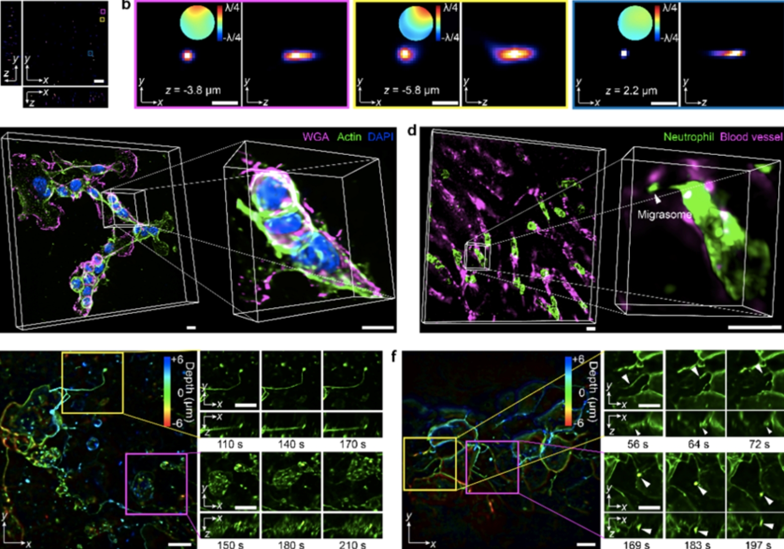
Long-term mesoscale imaging of 3D intercellular dynamics across a mammalian organ, Cell 2024
A miniaturized mesoscope for the large-scale single-neuron-resolved imaging of neuronal activity in freely behaving mice, Nature Biomedical Engineering 2024
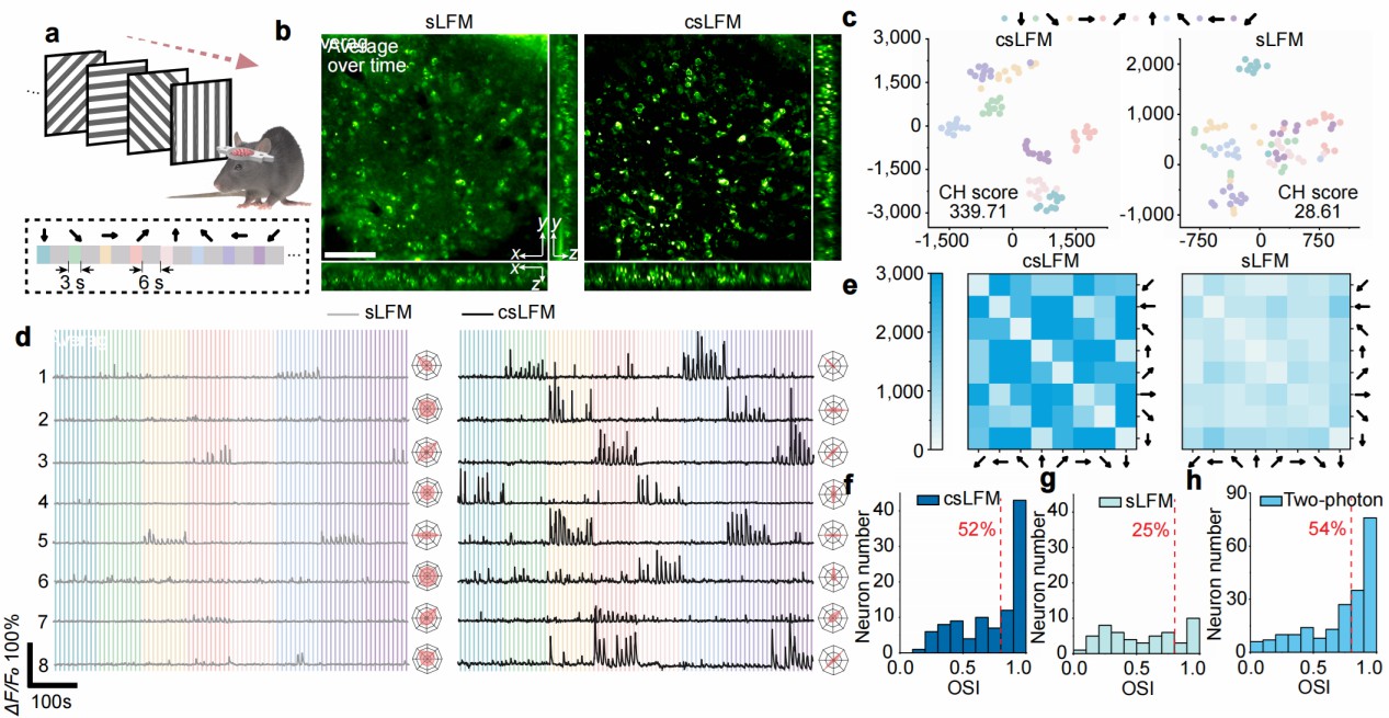
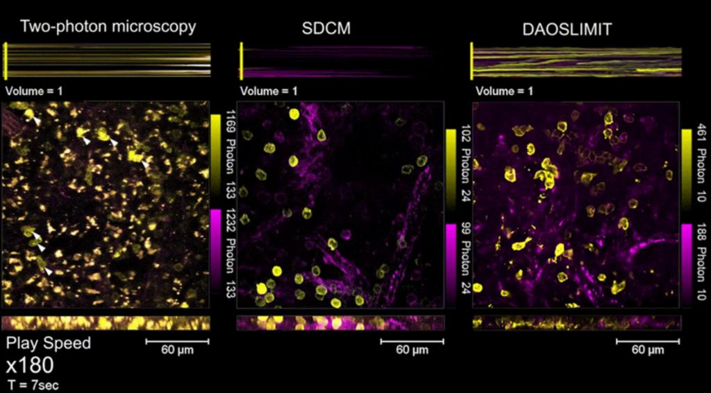
Long-term intravital subcellular imaging with confocal scanning light-field microscopy, Nature Biotechnology 2024
A Systematically Optimized Miniaturized Mesoscope (SOMM) for large-scale calcium imaging in freely moving mice, Nature Biomedical Engineering 2024
Multifocal fluorescence video-rate imaging of centimetre-wide arbitrarily shaped brain surfaces at micrometric resolution, Nature Biomedical Engineering 2024
Two-photon synthetic aperture microscopy for minimally invasive fast 3D imaging of native subcellular behaviors in deep tissue, Cell 2023
Multi-focus light-field microscopy for high-speed large-volume imaging, PhotoniX 2022
Iterative tomography with digital adaptive optics permits hour-long intravital observation of 3D subcellular dynamics at millisecond scale, Cell 2021
Video-rate imaging of biological dynamics at centimetre scale and micrometre resolution, Nature Photonics 2019
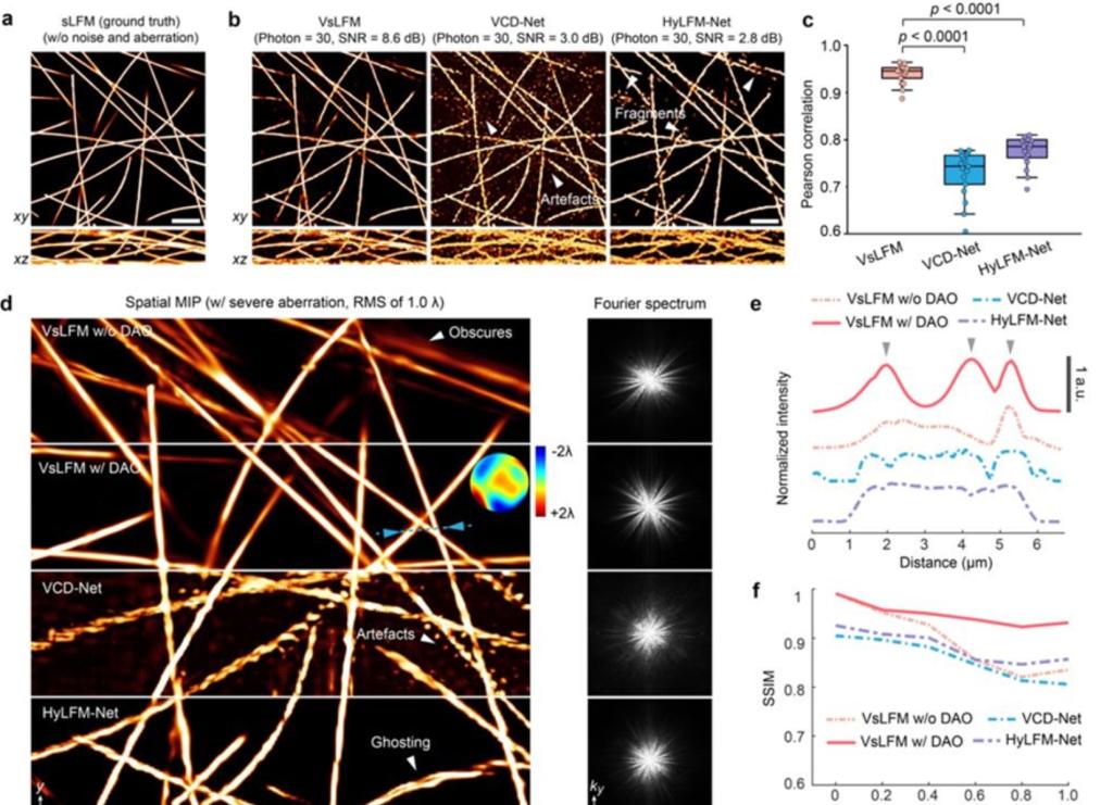
Virtual-scanning light-field microscopy for robust snapshot high-resolution volumetric imaging, Nature Methods 2017
 Mailbox:qhdai@tsinghua.edu.cn
 Address:3rd floor, Central Main Building, THU, Beijing
 Fax:+86 10 62773433


