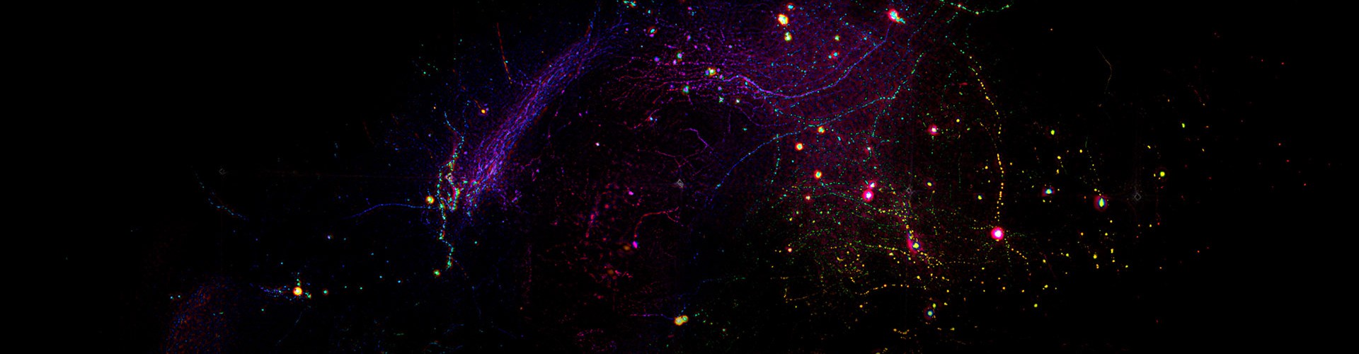
Fluorescence lifetime imaging microscopy (FLIM) measures the duration that a fluorescent molecule remains excited before radiative decay, providing quantitative information on molecular environments, interactions, and protein conformations. However, accurate lifetime determination conventionally requires repeated excitation and the accumulation of a large number of photons at each pixel. This photon requirement restricts the practical application of FLIM in fast, low-light, minimally phototoxic, and deep-tissue imaging.
To address this challenge, a team led by Qionghai Dai and Jiamin Wu from the Department of Automation, Tsinghua University, developed event-based first-photon fluorescence lifetime imaging microscopy (EFLIM), enabling high-speed FLIM in the single-photon regime.
Instead of constructing per-pixel photon-arrival histograms, EFLIM interprets each excitation event as a binary process: either no photon is detected, or a first-arrival photon is recorded with its precise arrival time. Based on this event-based representation, fluorescence lifetime estimation is reformulated as a self-supervised denoising problem. By exploiting sparse photon information from surrounding excitation events in both spatial and temporal domains, EFLIM recovers apparent mean-lifetime images without requiring high-photon reference images as supervisory labels.
Experimental results showed that EFLIM reduced the photon requirement by over two orders of magnitude compared with existing methods. Even under extremely low-light conditions, with an average photon count below 1 photon per pixel, EFLIM reliably reconstructed high-signal-to-noise fluorescence lifetime images.
The team further demonstrated EFLIM across multiple biomedical imaging scenarios:
Awake-mouse brain imaging: Under photon budgets of approximately 0.2–0.8 photons per pixel, EFLIM showed strong robustness to calcium-dependent and motion-induced intensity fluctuations, producing stable lifetime measurements at both the soma and single-pixel levels.
Live-cell calcium imaging: At approximately 0.2–0.5 photons per pixel, EFLIM recovered rapid intracellular lifetime changes and revealed temporal differences in calcium responses between subcellular regions within the same cell.
Mouse lymph-node imaging: EFLIM enabled multiplexed imaging of germinal-center B cells and follicular helper T cells within a single spectral channel, while capturing lymphocyte migration, direct cell–cell contacts, and putative vesicle-mediated contacts between T cells.
Human glioma tissue imaging: The team acquired 169 fields of view and generated a centimeter-scale, label-free lifetime map of a fixed human glioma tissue section. EFLIM required only 0.33 seconds per field of view, approximately one-tenth of the acquisition time used by the conventional method, while preserving tissue-scale and microstructural information and revealing lifetime heterogeneity across different tissue regions.
This work shifts the FLIM measurement paradigm from accumulating large numbers of photons to construct histograms toward fully exploiting each excitation event and first-arrival photon. By pushing the sensitivity limit of FLIM toward the single-photon level, EFLIM provides a new route for fast, low-light, minimally phototoxic, and deep-tissue imaging of dynamic molecular processes.
Associate Professor Jiamin Wu and Professor Qionghai Dai from the Department of Automation, Tsinghua University, are the co-corresponding authors. Doctoral candidate Yiliang Zhou from the Department of Automation, doctoral candidate Yihong Xiao from the College of AI, and postdoctoral researcher Jing Zhou from the Department of Automation are the co-first authors. The team has conducted sustained research in light-field microscopy, adaptive optics, and computational imaging, advancing their applications in the life sciences.

 Mailbox:qhdai@tsinghua.edu.cn
 Address:3rd floor, Central Main Building, THU, Beijing
 Fax:+86 10 62773433