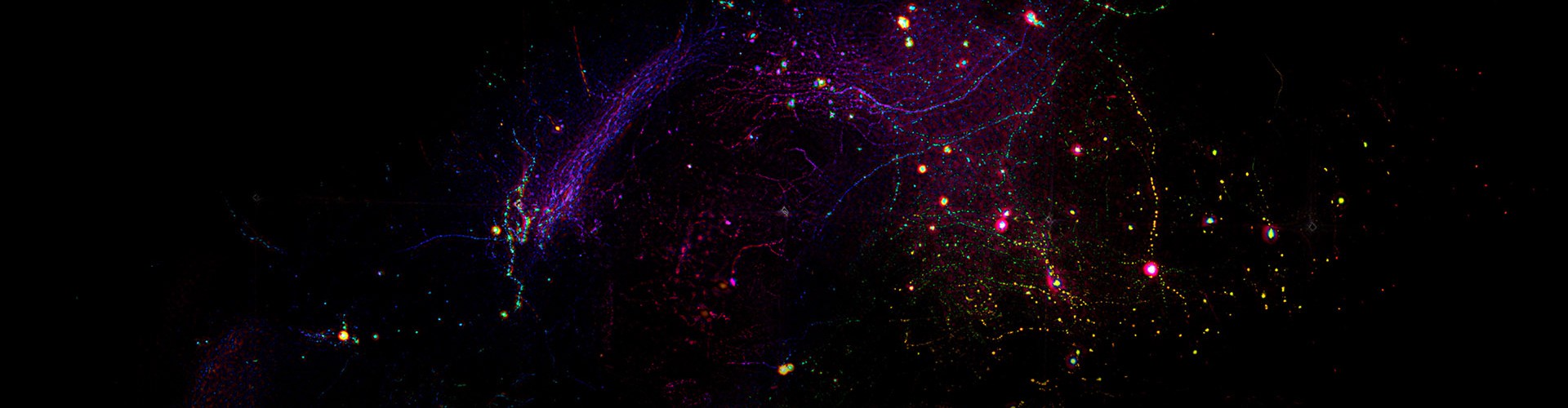
Within mammalian organisms, millions of immune cells and other functional cells continually undergo directed migration and coordinated interactions, collectively regulating a range of physio pathological processes including immune responses and inflammatory reactions. Over the past decade, although mesoscale intravital microscopy has made substantial progress, there remains a notable gap in imaging tools capable of simultaneously achieving subcellular resolution and mesoscale field of view in living specimens. Existing systems struggle to attain nanoscale resolving power across millimeter-scale fields, preventing researchers from directly tracking subcellular dynamics within large cell populations under pathological conditions. This limitation has severely constrained in-depth investigations into the immunomodulatory mechanisms and therapeutic efficacy of interventions such as acupuncture at the organismal level.
To address these challenges, a team lead by Dai Qionghai/Wu Jiamin from Tsinghua University and Wang Wei from the Department of Neurology, Tongji Hospital, Tongji Medical College, Huazhong University of Science and Technology, successfully developed a high-numerical-aperture mesoscale confocal volumetric microscope, designated RUSH3D-HR. The system integrates a 1.05 numerical aperture (NA) Kunlun objective lens, digital adaptive optical correction, and a confocal scanning light-field microscopy architecture, achieving a breakthrough in subcellular-resolution three-dimensional imaging across millimeter-scale fields of view, with a single-exposure imaging volume of 2.7 × 2.0 × 0.04 mm3. The imaging rate reaches up to 5 volume acquisitions per second, while maintaining low phototoxicity. The team specifically designed the Kunlun objective with a high lateral space–bandwidth product and paired it with a digital adaptive optics framework to overcome the aberration sensitivity inherent to high-NA systems. By adopting a scanning light-field microscopy configuration, they achieved parallel, high-speed, low-phototoxicity volumetric imaging; line-confocal technology was further employed to deliver robust optical sectioning and background rejection across a 40-μm axial range, effectively mitigating the shallow depth-of-field issue associated with high-NA optics. Moreover, to handle the massive data throughput of up to 70 giga voxels per second generated by mesoscale imaging, the team independently developed a deep learning–based parallel computational imaging pipeline that performs denoising, multi-point aberration correction, motion artifact removal, and three-dimensional reconstruction in an integrated workflow.
In system characterization, RUSH3D-HR achieved a lateral resolution of approximately 390 nm and an axial resolution of roughly 700 nm, with efficient processing capability for massive datasets—performance metrics substantially surpassing those of comparable technologies. The practicality and reliability of the system have been validated across multiple intravital imaging applications, including longitudinal tracking of mouse skin wound healing over several hours, observation of subcellular-level immune cell interactions in an acute liver failure model, and capture of transient neutrophil surge behavior in the spleen of mice with lipopolysaccharide-induced inflammation.
RUSH3D-HR possesses cross-scale observational capacity, enabling simultaneous visualization of dynamic processes ranging from organelles and single cells to whole-organ scales. Using this platform, the research team observed, for the first time in living splenic tissue, transient neutrophil aggregation events associated with inflammation. Furthermore, the team established this aggregation behavior as a mesoscale inflammatory imaging biomarker and systematically evaluated the anti-inflammatory effects of electroacupuncture stimulation at the ST36 acupoint.
The co-corresponding authors of this work are Professor Dai Qionghai, Associate Professor Wu Jiamin from the Department of Automation, Tsinghua University, and Professor Wang Wei from the Department of Neurology, Tongji Hospital, Tongji Medical College, Huazhong University of Science and Technology. Assistant Professor Lu Zhi from the Department of Psychological and Cognitive Sciences, Tsinghua University; Professor Wang Minghuan from the Department of Neurology, Tongji Hospital, Tongji Medical College, Huazhong University of Science and Technology; and doctoral candidate Chen Wentao from the Institute of Future Information Innovation, Fudan University, are co-first authors. The team has conducted sustained research in light-field microscopy, adaptive optics, and computational imaging, advancing their applications in life sciences.

 Mailbox:qhdai@tsinghua.edu.cn
 Address:3rd floor, Central Main Building, THU, Beijing
 Fax:+86 10 62773433