High-throughput recording of neural activity has greatly advanced our understanding of the spatiotemporal dynamics and functional organization of neurons1–3. However, other types of brain signals—such as the multi-regional dynamics of neurotransmitters and neuromodulators, as well as their spatiotemporal relationships with neuronal activity—remain poorly understood4. This lack of knowledge limits our systematic understanding of the functional roles of these chemical signals.
Taking the olfactory processing system of Drosophila as an example, although numerous neurotransmitters and neuromodulators, such as acetylcholine (ACh) and serotonin (5-HT), are known to play key roles, their stimulus-related spatiotemporal dynamics remain largely unknown. Moreover, functional overlaps among different neurotransmitters have been frequently observed in previous studies. Additionally, current investigations of olfactory representation in Drosophila have primarily focused on the early stages of olfactory processing, while our understanding of information representation in higher-order brain regions remains limited.
In recent years, rapid advances in fluorescent indicators for chemical signals and microscopy technologies have made it possible to image large-scale, high-throughput neuronal activity and neurochemical dynamics simultaneously—offering new opportunities to address these challenges.
On September 30th, our team published a paper in Nature Communications entitled “Prominent involvement of acetylcholine dynamics in stable olfactory representation across the Drosophila brain.” Using two-photon synthetic aperture microscope (2pSAM)5, they achieved two-hour-long, high-speed volumetric imaging of calcium and ACh6/5-HT7 signals across multiple brain regions of Drosophila. Through systematic information representation and network analyses, the study revealed the critical involvement of ACh dynamics in stable olfactory representation.
In this study, volumetric imaging was performed over a field of view measuring 458.7 μm × 458.7 μm × 100 μm, covering the entire central brain of Drosophila in the lateral dimension and approximately half of its thickness along the axial dimension. This configuration enabled the capture of neuronal activity across 43 distinct brain regions. Through genetic manipulation, the authors generated Drosophila lines in which all neurons across the whole brain simultaneously expressed calcium and ACh/5-HT indicators, allowing for concurrent imaging of neuronal activity and neurochemical dynamics across multiple brain regions.
Benefiting from the three-dimensional imaging capability and low phototoxicity of the 2pSAM system, the study achieved continuous volumetric recording for two hours at a volume rate of 30 Hz. During this period, the flies were presented with 180 odor stimuli, corresponding to three different odorants delivered in a randomized sequence. The multiple rounds of prolonged olfactory stimulation enabled analyzing olfactory representation and its temporal stability. By combining the recordings with the latest denoising algorithms8,9, the researchers extracted olfactory response signals with high signal-to-noise ratio comprising more than 200,000 voxels from each individual fly.
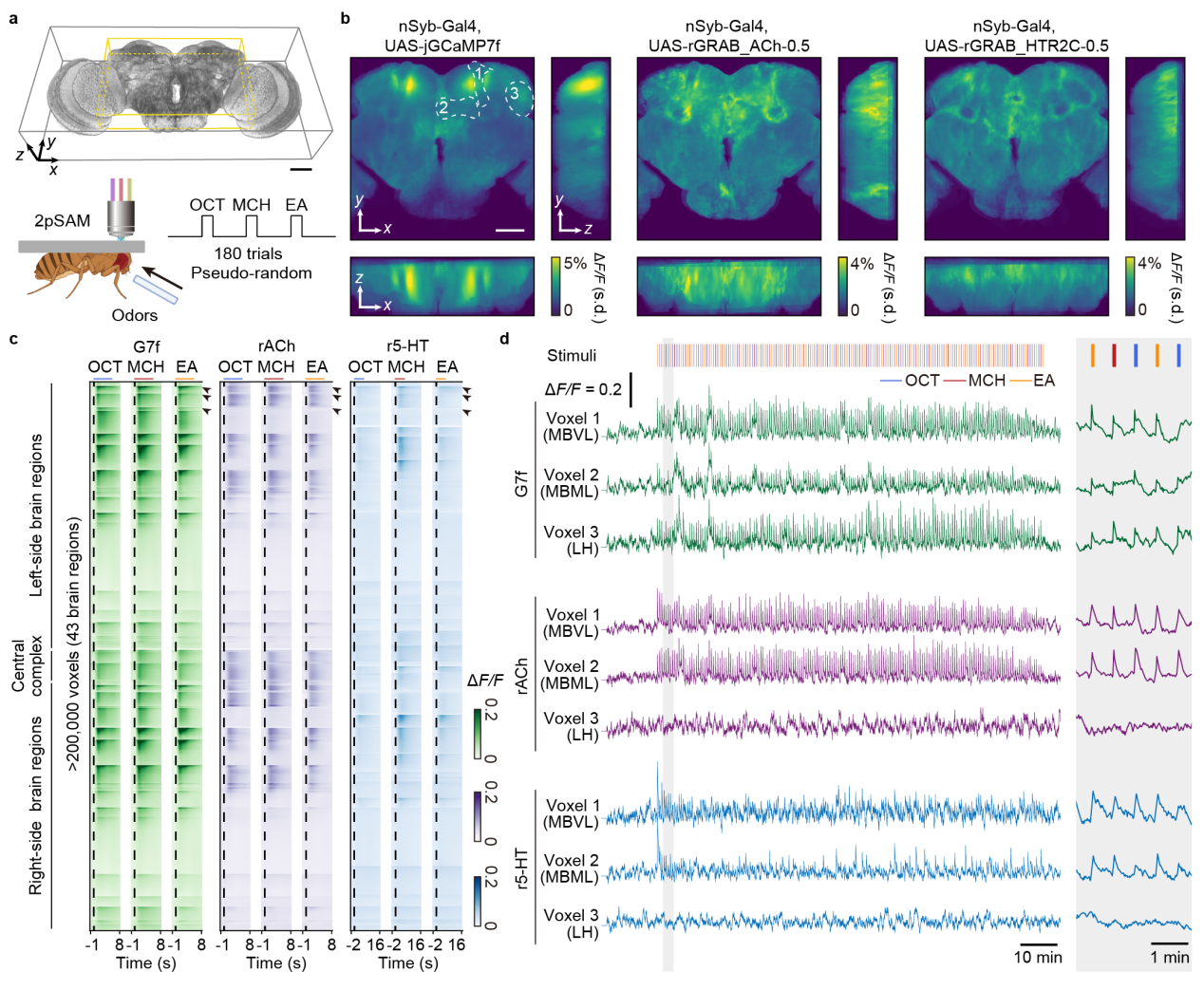
Figure 1. Volumetric imaging of neuronal activities and neurochemical dynamics across multiple regions in the Drosophila central brain by 2pSAM across 2 h.
Analyses of responsiveness, temporal response characteristics, odor classification accuracy, and functional connectivity across brain regions revealed substantial heterogeneity among different signals as well as across distinct brain areas. In terms of odor classification accuracy, calcium signals exhibited relatively high performance across multiple regions, with particularly high accuracy observed in the lateral horn. In contrast, regions showing high accuracy for ACh and 5-HT signals were more spatially localized. Furthermore, by integrating neural activity information from multiple brain regions, the study constructed low-dimensional manifolds representing olfactory information for all three signal types, and statistically compared the characteristics of manifolds. Overall, the representational accuracy of calcium and ACh signals was comparable and both exceeded that of 5-HT. Notably, calcium signals demonstrated clearer odor preference encoding within the low-dimensional manifold—showing distinct manifold characteristics for appetitive and aversive odors, while odors sharing the same valence were less separable. In contrast, although ACh signals could distinguish among the three odors, their encoding of odor valence was less pronounced. The study also identified spatial ensembles representing the three odors under each signal type.
Moreover, integrating calcium and ACh signals resulted in a further improvement in odor classification accuracy, both within local regions and at the level of the multi-region low-dimensional manifold. By comparing voxel-level functional connectivity within specific brain regions, the authors found complementary functional coupling between calcium and ACh signals in regions showing accuracy enhancement. In contrast, combining calcium and 5-HT signals did not yield similar improvements in accuracy or functional connectivity complementarity.
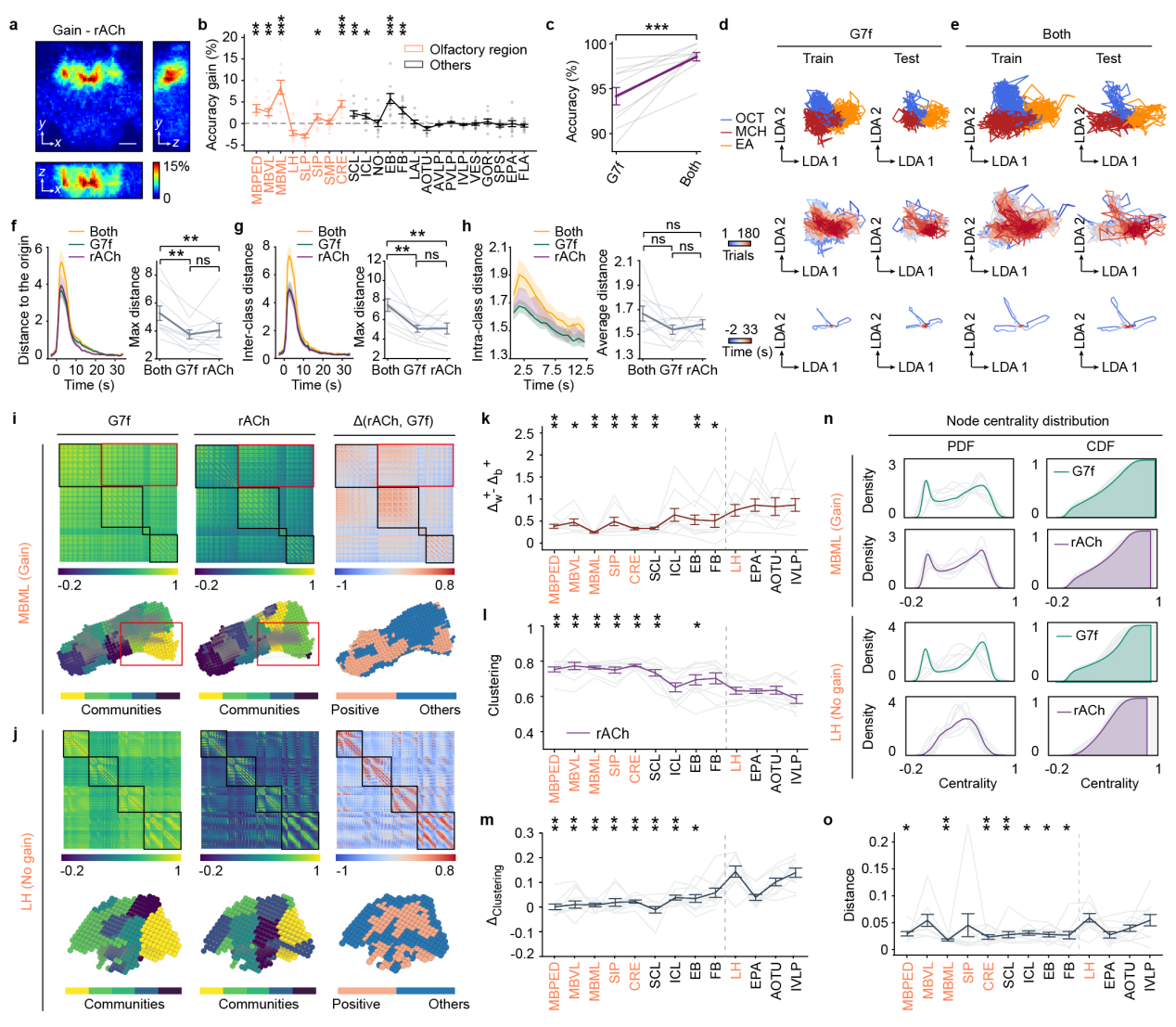
Figure 2. Integration of ACh dynamics improves the odor identity representation by neuronal activity.
Finally, the study analyzed changes in the low-dimensional manifolds of odor representations and functional connectivity across 180 odor stimulation trials. The 180 trials were evenly divided into four sequential stages, and the relevant metrics were quantified for each stage. The analysis revealed that ACh dynamics remained the most stable across stages, whereas calcium signals exhibited progressive changes over time, and 5-HT signals showed a notable attenuation primarily during the initial stages. All three signal types displayed a reduction in within-class distances between the first and second stages of stimulation, suggesting potential learning or adaptation effects underlying the evolving odor representations.
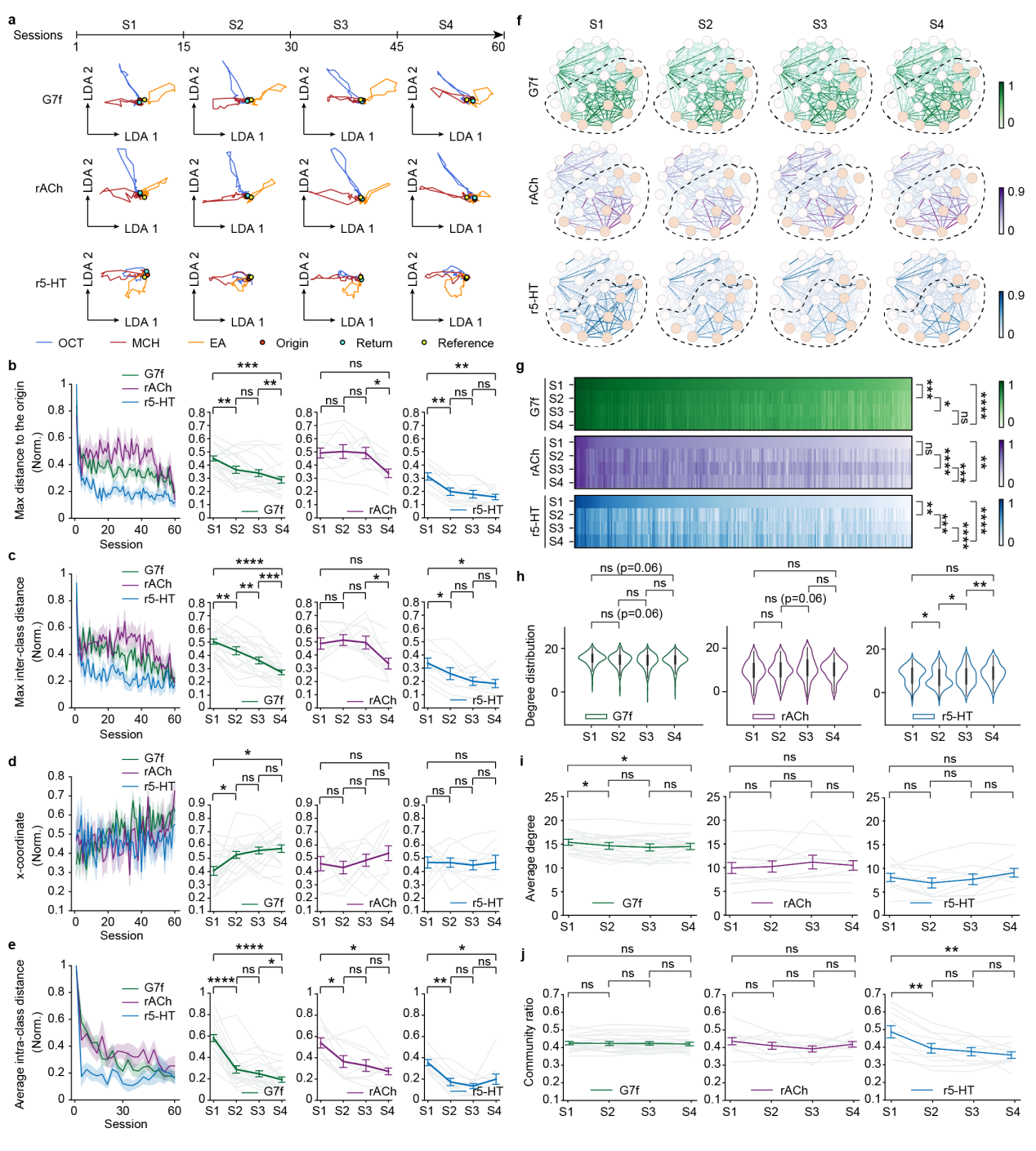
Figure 3. ACh signals exhibit greater temporal stability in odor representation and the functional connectivity network than calcium and 5-HT.
In summary, this study employed multi-region, multi-signal recording combined with systematic analysis to uncover the critical role of ACh dynamics in establishing stable olfactory representations across the Drosophila brain. These findings highlight the importance of integrating multi-signal observations and analyses for a comprehensive understanding of neural information representation. The full dataset has been publicly released to facilitate further research and collaboration within the scientific community.
Jiaqi Fan, a Ph.D candidate in our lab, currently a Postdoctoral Fellow in City University of Hong Kong; Yuling Wang, a master student in our lab, currently at Carnegie Mellon University; and Lingbo Li, a postdoctoral researcher in our lab are co–first authors of the paper. Prof. Qionghai Dai, Prof. Lu Fang, and Prof. Jiamin Wu are co–corresponding authors. Additional contributors include Prof. Yulong Li, Dr. Fei Deng, and Guochuan Li from the School of Life Sciences, Peking University; Dr. Jing He, Dr. Zhifeng Zhao, Ph.D. candidates Yiliang Zhou, Jiayin Zhao, and Yixin Hu from the Department of Automation, Tsinghua University; Asst. Prof. Xinyang Li from College of AI, Tsinghua University; and Dr. Ning Huang from the School of Life Sciences, Tsinghua University. This work was supported by the National Natural Science Foundation of China (NSFC).
Link:https://www.nature.com/articles/s41467-025-63823-2
References:
1. Steinmetz, N. A., Zatka-Haas, P., Carandini, M. & Harris, K. D. Distributed coding of choice, action and engagement across the mouse brain. Nature 576, 266–273 (2019).
2. Ebrahimi, S. et al. Emergent reliability in sensory cortical coding and inter-area communication. Nature 605, 713–721 (2022).
3. Brezovec, B. E. et al. Mapping the neural dynamics of locomotion across the Drosophila brain. Current Biology 34, 710-726.e4 (2024).
4. Urai, A. E., Doiron, B., Leifer, A. M. & Churchland, A. K. Large-scale neural recordings call for new insights to link brain and behavior. Nat Neurosci 25, 11–19 (2022).
5. Zhao, Z. et al. Two-photon synthetic aperture microscopy for minimally invasive fast 3D imaging of native subcellular behaviors in deep tissue. Cell 186, 2475-2491.e22 (2023).
6. Jing, M. et al. An optimized acetylcholine sensor for monitoring in vivo cholinergic activity. Nat Methods 17, 1139–1146 (2020).
7. Deng, F. et al. Improved green and red GRAB sensors for monitoring spatiotemporal serotonin release in vivo. Nat Methods 21, 692–702 (2024).
8. Li, X. et al. Real-time denoising enables high-sensitivity fluorescence time-lapse imaging beyond the shot-noise limit. Nat Biotechnol 41, 282–292 (2023).
9. Li, X. et al. Spatial redundancy transformer for self-supervised fluorescence image denoising. Nat Comput Sci 3, 1067–1080 (2023).
 Mailbox:qhdai@tsinghua.edu.cn
 Address:3rd floor, Central Main Building, THU, Beijing
 Fax:+86 10 62773433