Two-photon microscopy is indispensable for in vivo observation of deep, scattering tissues, with its superior penetration depth compared to single-photon imaging being widely recognized in life sciences and medical research. However, traditional point-scanning two-photon imaging fundamentally limits imaging throughput and three-dimensional perception speed, making it highly susceptible to interference from complex in vivo environments. Additionally, the high instantaneous light intensity at the excitation point causes persistent nonlinear photodamage to biological samples, severely restricting the duration of high-speed three-dimensional imaging and hindering advancements in pathology, immunology, and neuroscience.
Addressing these challenges, on May 12, 2023, Qionghai Dai, Jiamin Wu, and Hai Qi from Tsinghua University published a study titled ‘Two-photon synthetic aperture microscopy for minimally invasive fast 3D imaging of native subcellular behaviors in deep tissue’ in Cell[1]. They introduced a spatially constrained multi-angle diffraction encoding method to achieve incoherent light aperture synthesis, establishing two-photon synthetic aperture microscopy (2pSAM). This technique transforms ‘points’ into ‘needles’, using multi-angle needle-like beam scanning to achieve high-speed three-dimensional perception while reducing two-photon imaging phototoxicity by over 1,000 times. Integrating the digital adaptive optics (DAO)[2] framework proposed by Qionghai Dai's team in 2021, 2pSAM offers high-speed multi-region aberration correction, maintaining near-diffraction-limited spatial resolution even in complex in vivo environments and further enhancing the penetration depth of traditional two-photon imaging. Consequently, 2pSAM enables non-invasive observation of large-scale subcellular dynamics in deep scattering tissues of mammals, extending millisecond-scale continuous three-dimensional observation duration from minutes to tens of hours, thereby opening new avenues for systematically studying large-scale cellular interactions under different physiological and pathological states. Using 2pSAM, the interdisciplinary research team observed a series of novel phenomena in vivo in mice, including multicellular interactions around brain tissue following acute brain injury, neuronal representation stability and functional diversity in response to visual stimuli during long-term continuous observation, and the first complete high-speed recording of germinal center formation in lymph nodes during immune responses, thus providing new insights for research in pathology, neuroscience, and immunology.

Traditional two-photon microscopy uses ‘point scanning’ to scan three-dimensional samples, similar to confocal fluorescence microscopy. Due to the nonlinear effects of two-photon imaging, it achieves several times the penetration depth of single-photon imaging. For example, two-photon microscopy can reach a maximum penetration depth of 1 mm in the mouse cortex. However, this point-scanning method severely limits three-dimensional imaging speed and data throughput, and the high instantaneous light intensity at the focus point poses a significant risk of nonlinear photodamage. 2pSAM employs an axial depth-extended ‘needle scanning’ scheme, achieving multi-angle projections of three-dimensional sample information by varying the inclination angles of needle-like beams, similar to CT for rapid three-dimensional imaging. Inspired by synthetic aperture methods in radar imaging, spatial diffraction encoding constraints introduced at the image plane enable incoherent light aperture synthesis, combining multi-angle information into high spatial resolution corresponding to a large numerical aperture. Additionally, leveraging the spatiotemporal continuity priors of samples effectively avoids time resolution loss due to angular scanning. This novel computational two-photon imaging framework retains the deep tissue penetration capability of two-photon microscopy while increasing effective imaging throughput by over three orders of magnitude.

Figure 1. Schematic of Two-photon Synthetic Aperture Microscopy (2pSAM) system
Moreover, optical aberrations induced by samples significantly degrade imaging resolution and signal-to-noise ratio, with this degradation becoming more pronounced as imaging depth increases. Current hardware adaptive optics techniques in two-photon imaging face challenges such as system complexity, high costs, limited effective correction fields, and slow correction speed in large field-of-view multi-region corrections. 2pSAM acquires ultra-fine four-dimensional spatial angular light field data through excitation light encoding, enabling the use of DAO to decouple signal acquisition and adaptive aberration correction without adding extra wavefront sensors or spatial modulators to the optical system. This allows for large-scale multi-region adaptive optics correction during post-processing, significantly improving spatial resolution and signal-to-noise ratio in complex imaging environments.

Figure 2. Comparison of results between 2pSAM combined with DAO and traditional two-photon microscopy (TPM) under complex imaging conditions. From left to right: normal conditions, mismatched objective correction ring, water objective without immersion water, and imaging with added scattering tape between the objective and sample
Extended laser exposure causes significant phototoxicity to live samples. The research team found that traditional two-photon imaging, using femtosecond laser excitation and high NA focusing, generates substantial instantaneous light intensity at the sample, with resulting nonlinear phototoxicity being severely underestimated in the past. This accumulative damage affects normal cell states during long-term imaging. In contrast, 2pSAM transforms points into needles, reducing instantaneous peak power by 1,000 times while maintaining the same fluorescence excitation efficiency, effectively addressing nonlinear photodamage. This significantly reduces fluorophore photobleaching; for instance, while traditional two-photon can only capture tens of three-dimensional volumes before signal decay, 2pSAM can continuously capture hundreds of thousands of three-dimensional volumes without noticeable signal attenuation. Furthermore, continuous imaging tests on microglia and neutrophils during brain injury in mouse cortex showed that traditional two-photon imaging, even at lower light intensities, leads to significant cell apoptosis after half an hour of continuous imaging, whereas cells imaged with 2pSAM maintained normal phenotypes with no significant differences compared to control groups. This series of in vivo and in vitro experiments demonstrated that 2pSAM reduces phototoxicity by over three orders of magnitude compared to traditional two-photon imaging, paving the way for long-term high-speed in vivo tissue imaging.

Figure 3. Imaging of cortical immune cells after acute window injury in mouse brain, comparing photobleaching between TPM (left) and 2pSAM (right)

Figure 4. Continuous imaging of ex vivo B cells (GFP, blue channel) using PI to label apoptosis (red channel), comparing phototoxicity between TPM (left) and 2pSAM (right)
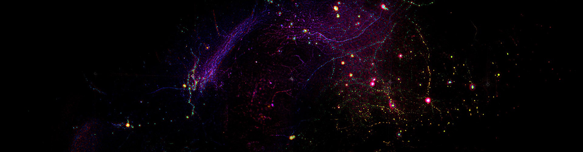
Germinal centers (GCs) are dynamic regions in secondary lymphoid organs where antigen-activated B cells congregate, playing a crucial role in immune responses. However, due to the randomness of GC formation and the sensitivity of immune cells to photodamage, the complete process of GC formation has never been clearly recorded over long durations at high speeds. Utilizing 2pSAM, researchers achieved the first clear and complete observation of the entire GC formation process during an immune response. Researchers labeled B cells in the inguinal lymph nodes of mice and continuously tracked the dynamic behavior of B cells under large field-of-view conditions 90 to 110 hours post-immunization, revealing that B cell proliferation drives GC formation, supplemented by the aggregation of surrounding mature B cells. Given the imaging duration of over ten hours, the lymph nodes undergo significant deformation. 2pSAM’s multi-angle information allows for real-time axial focus position feedback, achieving automatic focus and effectively avoiding sample drift during long-term imaging.

Figure 5. Complete observation and recording of germinal center formation in the inguinal lymph nodes of mice post-immune response
Researchers further used 2pSAM to observe cellular dynamics in the cortical tissues of mice with traumatic brain injury (TBI) and GCaMP transgenic mice undergoing visual stripe stimulation. After thinning the skull in the injured area of TBI mice, they observed interactions between peripheral neutrophils and astrocytes in the injured region, such as directed formation of migrasomes through direct contact for substance and information transmission. In GCaMP transgenic mice, two weeks post-craniotomy, visual stripe stimulation further confirmed the sustained and stable expression of calcium signals in visual cortex neurons in response to different directional stripes over several hours. Additionally, long-term functional data mining revealed various neuron response types at the single-cell level, reflecting neuronal functional diversity. These phenomena are highly challenging for traditional two-photon microscopy, particularly due to the phototoxicity causing abnormal cell responses, such as declining neuron response strength during long-term imaging.
Article link: https://www.cell.com/cell/fulltext/S0092-8674(23)00412-9
References:
[1] Zhao, Zhifeng, et al. Two-photon synthetic aperture microscopy for minimally invasive fast 3D imaging of native subcellular behaviors in deep tissue. Cell (2023).
[2] Wu, Jiamin, et al. Iterative tomography with digital adaptive optics permits hour-long intravital observation of 3D subcellular dynamics at millisecond scale. Cell 184.12 (2021): 3318-3332.
 Mailbox:qhdai@tsinghua.edu.cn
 Address:3rd floor, Central Main Building, THU, Beijing
 Fax:+86 10 62773433