Burst-suppression is a characteristic brain activity pattern commonly observed during deep anesthesia, coma, hypothermia, and certain pathological states of unconsciousness. It is marked by alternating episodes of high-amplitude electrical bursts and periods of strongly suppressed activity. For decades, this phenomenon has been primarily observed through macroscopic electroencephalographic signals and has often been interpreted as a cortex-wide alternation between “global activation” and “global silence.”
Yet important questions remain: Does a “flat” electroencephalographic signal truly mean that neurons have completely stopped firing? Does a burst reflect simultaneous activation across the entire cortex? Answering these questions requires placing macroscopic electrical activity and tens of thousands of single-neuron signals onto the same spatiotemporal map.
To address this cross-scale challenge, Prof. Qionghai Dai and Prof. Jiamin Wu from the Department of Automation at Tsinghua University, in collaboration with Prof. Xiaochuan Dai from the School of Biomedical Engineering at Tsinghua University, developed a cortex-wide optical-electrical recording system named CODE — Cortex-wide Optical-electrical Dual-modal Explorer. Their study, titled “Dynamic neuronal ensembles encode burst-suppression revealed by cortex-wide optical-electrical interfaces,” was published in Nature Communications on April 29.
The CODE system integrates transparent electrocorticography arrays with an ultra-large-field, single-cell-resolution calcium imaging platform, enabling simultaneous recording of large-scale neuronal activity and multi-region electrophysiological signals.
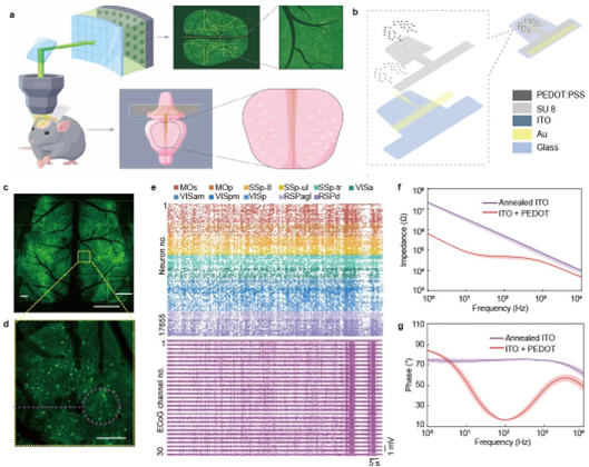
Figure 1. Cortex-wide optical-electrical recording system CODE
Using CODE, the team simultaneously recorded cortex-wide ECoG signals and calcium activity from tens of thousands of neurons in mice under isoflurane anesthesia. As anesthesia deepened, typical burst-suppression rhythms emerged in the ECoG signals, accompanied by pronounced state-dependent changes in neuronal calcium activity.
Further analysis revealed two distinct neuronal populations in the cortex: one population was more active during burst phases, while the other was more active during suppression phases. This finding indicates that “suppression” in ECoG does not mean that neurons are completely silent. Instead, neuronal activity persists in a more dispersed and asynchronous form. The researchers identified burst-related and suppression-related neuronal populations that alternated with the burst-suppression rhythm, demonstrating that this brain state is organized by distinct neuronal ensemble structures.
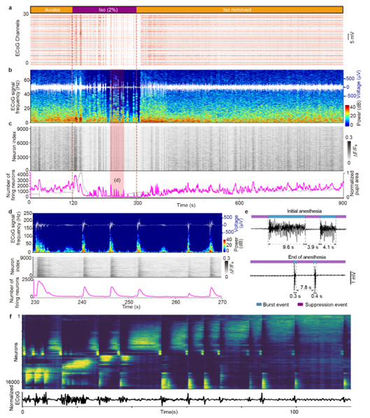
Figure 2. Alternating activation of two neuronal populations during burst-suppression
The team further tracked the dynamic evolution of these two neuronal populations throughout anesthesia. The results showed that burst-related and suppression-related neurons were not fixed groups. As anesthesia progressed, burst duration shortened and the number of participating neurons decreased, while suppression periods gradually lengthened and the number of suppression-related neurons also declined. Each event involved both newly recruited neurons and neurons that had participated in previous events, suggesting that burst-suppression is a continuously evolving process of neuronal ensemble reorganization.
In terms of temporal structure and network organization, burst-related neurons were typically recruited rapidly and synchronously during the early phase of bursts, forming stronger functional connections. In contrast, suppression-related neurons were recruited later and in a more dispersed manner, with weaker connection strength. These distinct ensemble dynamics together shaped the differences between burst and suppression waveforms observed in macroscopic ECoG signals.
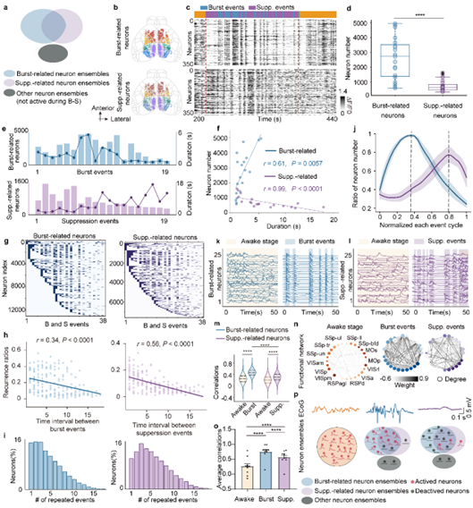
Figure 3. Dynamic recruitment and functional connectivity of neuronal ensembles during burst-suppression
The study also found that the transition from suppression to burst did not occur as an instantaneous, globally synchronized event. Instead, it followed a clear cortical spatiotemporal structure. Multi-channel ECoG analysis showed that burst activity usually emerged first in bilateral lateral sensory cortices and then propagated toward medial and anterior motor-related regions.
Single-cell calcium activity closely mirrored this process. Neuronal activity in lateral sensory cortices was more dispersed, whereas activity peaks in medial motor-related regions were sharper and more tightly aligned with burst onset. Cross-modal correlation analysis between ECoG and calcium signals further showed that synchronization between electrical and neuronal activity gradually increased during propagation, revealing a process of “synchronization during propagation.”
Together, these findings show that burst-suppression is not a uniform, passive process of cortex-wide silence. Rather, it is a complex brain state shaped by dynamic neuronal ensemble recruitment, changes in functional connectivity, and coordinated spatiotemporal organization across cortical regions. By aligning large-scale single-neuron calcium imaging with multi-region ECoG recording on the same timeline, CODE provides a powerful cross-scale platform for investigating brain state transitions in anesthesia, sleep, coma, epilepsy, and related conditions.
Authors and affiliations:
Academician Qionghai Dai, Associate Professor Jiamin Wu, Associate Prof. Xiaochuan Dai, Associate Research Fellow Guihua Xiao from Tsinghua University, are the co-corresponding authors of this paper. Guihua Xiao, PhD student Mo Yang, and Postdoctoral Researcher Lingbo Li, are the co-first authors. Collaborators also include Professor Bo Hong from Tsinghua University and Professor Ting Lei from Peking University.
Original article: https://www.nature.com/articles/s41467-026-72454-0
 Mailbox:qhdai@tsinghua.edu.cn
 Address:3rd floor, Central Main Building, THU, Beijing
 Fax:+86 10 62773433